


Varicose veins of the lower extremities are characterized by dilation of the superficial veins of the legs, which is accompanied by violation of blood flow and failure of the valves. As a result, the length and diameter of the vessels increase, acquiring a serpentine, cylindrical, or saccular appearance, although the listed deformities are also mixed.
Characteristics of the venous system
The formation and formation of varicose veins is directly related to the venous system of the legs, which consists of:
- saphenous veins: small and large;
- deep veins (in the lower leg and thigh);
- perforating vessels, which are the connecting elements of the two previous systems.
Normally, 90% of the blood is transported deep in the veins and the remaining 10% superficially to the lower extremities. When it returns to the side of the heart, this mechanism is supported by valves in the vessel wall. When the next part of the blood arrives, they collide to prevent it from moving from top to bottom under the influence of gravity. Muscle contractions continue to deliver blood to the heart, allowing normal blood flow.
If a person is in a vertical position for a long time, blood stagnation can develop, which increases the pressure in the veins and causes them to increase in diameter. This process causes the valve leaflets to close incompletely, causing the flow of blood to be disrupted by the reverse flow from the heart - reflux.
It is most likely to affect the deep vein valves as they carry the largest amount of blood and therefore experience the greatest load. To reduce the high pressure in them, some of the blood is transported by perforated veins to the superficial veins, which were not originally intended for large volumes. Such loading of the walls of the veins leads to their dilation and varicose veins.
At the same time, the blood enters the deep veins without stopping, but due to the violation of their functions and the normal activity of the valve leaflets of the perforated veins, the blood is redistributed to the superficial vessels. As a result, chronic varicose veins develop over time, accompanied by painful feelings, edema, and trophic ulcers.
Causes of the disease
Previously, one of the main causes of varicose veins was called hereditary factor, but today this theory has been refuted. Of course, it is possible to trace the common manifestations of the disease in some families, but this is more due to the peculiarities of the life lived in the family: eating culture, passive rest, sedentary work and the like.
Varicose vein formation is based on the presence of reflux in the venous system when blood circulates in opposite directions through the veins. Further transport of blood from deep veins to superficial veins is possible due to the congenital or acquired degenerative pathology of the valve device. This causes superficial blood vessels to become supersaturated and tense when venous nodules form.
One of the main causes of varicose veins is an unhealthy diet, which in some cases leads to obesity. Such people exercise little, consume mainly highly processed foods, and the proportion of plant fibers in the diet is minimized. After all, they are involved in strengthening the walls of veins and blood vessels and prevent prolonged chronic constipation, which greatly increases intra-abdominal pressure and thus provokes varicose veins. It should be noted that an increase in body weight of more than 20% increases the risk of the disease fivefold.
The main provocative factor for women is wearing a child, while the risk of varicose veins increases with each subsequent pregnancy. Severe weight gain and an enlarged uterus put a heavy strain on stagnant legs. This situation is exacerbated by the ever-increasing intra-abdominal pressure and the effect of the hormone progesterone, which affects the condition of the elastic fibers in the vessel wall.
Other factors that cause varicose veins in the lower extremities include:
- sedentary lifestyle, standing upright during the day (such as hairdressers), long flights or long journeys. All this leads to stagnant processes in the lower extremities when blood accumulates in the superficial veins and is poorly transported to the heart;
- occasionally increases the risk of developing varicose veins in women wearing uncomfortable, tight shoes, especially in high-heeled models;
- corsets and tight underwear compress the inguinal veins and increase intra-abdominal pressure, which is a direct prerequisite for varicose veins;
- hypertension;
- smoking, which indirectly leads to thinning of the vessel wall.
Classification of the disease
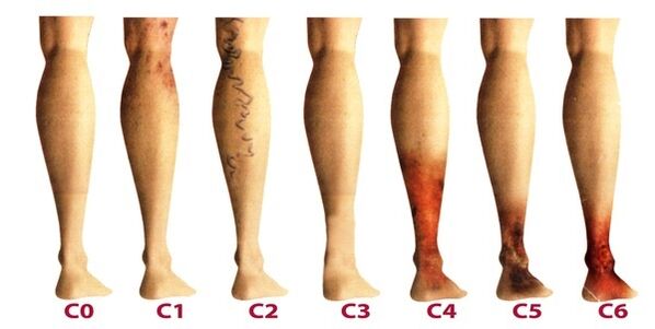
The varicose veins of the lower extremities are classified according to the prevalence and localization of venous lesions and the presence of abnormal reflux, which are characterized by harmful blood outflow. There are 4 forms of varicose veins:
- intracutaneous and subcutaneous varicose veins (segmental) without pathological outflow of venous blood;
- segmental varicose veins when reflux occurs through perforating or superficial veins;
- a common form of varicose veins in which reflux occurs simultaneously through the perforating and superficial veins;
- the varicose veins are characterized by deep vein reflux.
Once the varicose veins in the lower extremities become chronic, it considers three stages of phlebology:
- Transient edema that occurs intermittently in the background of "heavy legs" syndrome.
- Persistent, persistent edema. Hyperpigmentation and eczema may occur.
- Trophic venous ulcer.
The latter degree is the most difficult to treat as it requires prior removal of the inflammation and healing of the skin tissues.
Stages and symptoms
The disease develops very slowly, sometimes taking more than a dozen years before the onset of symptoms forces the patient to seek advice from a phlebologist. Manifestations in the early stages of varicose veins are often attributed to fatigue, age, or other causes. In order to take full account of the symptoms of the disease, its manifestations are classified according to the stages of the varicose veins:
- The first stage begins to manifest itself more often at a young age - after 20 years, when the feeling of heaviness in the legs occurs, edema may appear, which completely disappears overnight. Inside the lower leg, an enlarged vein is seen, manifested by a bumpy protrusion of the skin. At this stage, many people notice the little poker. Symptoms are usually mild and rarely get the attention they deserve.
- The second stage is characterized by an increase in the external manifestation of the dilated vein. The disease is already developing in the background of the abnormal work of the venous valves, so the size of the saphenous veins is noticeably increasing and their prolongation can be observed. More often there is difficulty and burns in the legs, they quickly get tired of long walks.
- The disease is already becoming chronic due to a constant imbalance in venous blood outflow. In the evenings, patients suffer from edema close to the ankles, which can be very intense. There is difficulty in the legs, cramps may occur at night.
- In the absence of treatment in the previous stages, chronic insufficiency of the venous system negatively affects the metabolic processes of the skin, especially the areas of the lower leg. Near the ankle you can see darkening of the skin - hyperpigmentation, thickens and inflames over time. The condition described is called lipodermatosclerosis. If you do not start therapy for the venous system at this time, trophic ulcers will soon begin to form.
- The fifth stage is accompanied by a number of trophic ulcers, some of which heal periodically with the formation of scars.
- Extensive ulcers open in the zone of long-standing trophic disorders. This condition requires urgent active therapy to treat both the varicose vein and the skin ulcer.
Diagnostics
External examination of the lower limbs in the vertical and horizontal position of the body, palpation of the veins and preliminary assessment of the stage of the disease. The patient is sent for a general blood test, which allows him to study the picture of the disease more thoroughly:
- the tendency to thrombosis is reflected at the platelet level;
- hemoglobin levels as well as red blood cell counts indicate the degree of blood clotting;
- elevated leukocyte levels can be used to judge inflammation, which helps to diagnose thrombophlebitis more quickly.
Be sure to look into the venous system of the legs, for which there are several methods:
- ultrasound dopplerography - USDG;
- phlebography;
- CT phlebography;
- duplex angioscanning - USAS;
- phleboscintiography;
- photoplasmography;
- phlebomanometry and the like.
In practice, patients are prescribed USAS and USG more often because they help to fully study the venous system of the legs and identify degenerative areas. Other methods may be prescribed if the ultrasound did not give a complete picture of the disease. Complications of some of these methods may include venous thrombosis, catheter perforation of the vessel wall, and allergy to the contrast agent. Consider the most commonly used techniques in phlebology:
- USAS allows the assessment of anatomical, hemodynamic, and functional pathologies of the venous bed. The data obtained is subjected to computer processing, after which the model of the venous system can be viewed on video or printed on paper.
- Doppler ultrasound determines with high accuracy the permeability of superficial and deep veins, the speed of blood flow. Doppler ultrasound allows the operation of the valve device to be assessed.
After extensive diagnosis, the physician prepares the patient’s phlebocard, which allows the damaged segments, extent, and length of the venous system to be determined. We then select an appropriate treatment.
Treatment
They are performed comprehensively and are determined based on symptoms, the rate of disease development, and the results of the study. In the initial phase, conservative therapy is prescribed, consisting of:
- Drug treatment if a group of drugs is prescribed:
- antiprotectors and phlebotonics;
- anticoagulants;
- disgregants
- topical preparations (ointments, gels);
- anti-inflammatory drugs.
- Flexible compression using a compression stocking or bandage (rarely). It allows the administration of muscle compression, prevents stagnant processes, improves blood flow through blood vessels. Wearing such underwear has the effect of artificially maintaining the tone of the vessel.
- Physiotherapy methods, among which the best treatment results were demonstrated by electrophoresis, diadynamic currents, laser radiation and magnetic field.
- Physical activity that can only be done in compression underwear (excluding swimming) is feasible. Cycling, swimming, jogging are recommended. The phlebologist chooses a series of individual exercises for the lower extremities that train the vessels of the legs every day.
In addition, we recommend that patients perform contrasting five-minute procedures in the shower every night, alternating between warm and cold. Such manipulations improve blood flow and tone the blood vessels.
At the beginning of treatment, it is important to determine the factor that provokes the disease in order to effectively influence it. And at-risk patients should visit a pre-phlebologist every 2 years and have an ultrasound scan of the veins in the leg.
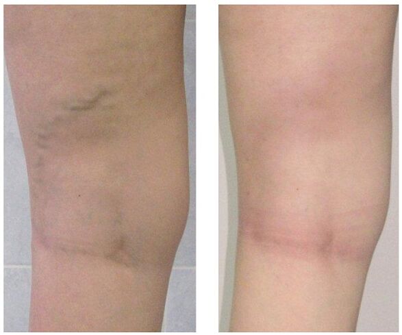
If conservative treatment does not produce results, or if the varicose vein is observed at an advanced stage, surgery is used. Today, varicose veins can be completely cured thanks to the following methods:
- Phlebectomy. The essence of the surgery is to remove the main trunk of the superficial vein to stop abnormal discharge of blood. Perforating veins are often ligated for the same purpose.
- Sclerotherapy. This consists of inserting a sclerosant into the affected area of the vein, leading to the connection of its walls. Recently, foam sclerosants have been actively used according to the technology for the same purposes. Blood flow through the defective area is stopped and the cosmetic defect in the form of protruding nodules is eliminated. After such an intervention, no scars remain, all manipulations are performed on an outpatient basis without any subsequent inpatient stay. But sclerotherapy is only used to fuse small branches of venous strains.
- Laser coagulation. A laser beam heats up the marked section of the vein, the walls of which stick together and stop the flow of blood through it. But this technique is only recommended for vessels with an expansion diameter of less than one centimeter.
Prevention
Preventive measures may be primary to prevent the development of varicose veins and secondary when it is necessary to reduce the risk of relapse after surgery or to prevent the progression of the disease. Useful tips:
- lead an active lifestyle without putting a heavy strain on your feet: swimming, walking, cycling;
- watch your weight;
- hold both feet more often;
- do not wear tight underwear and heels above 4 centimeters;
- use an orthopedic insole;
- take a contrast shower;
- perform five minutes of preventive leg exercises a day;
- wear compression stockings for long walks.
If you notice the slightest suspicion of varicose veins - prominent lumps on the legs, swelling, difficulty, do not delay the visit to the phlebologist. In fact, over time, this insidious disease can cause a plethora of complications, including thrombophlebitis and thrombosis.